


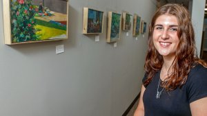


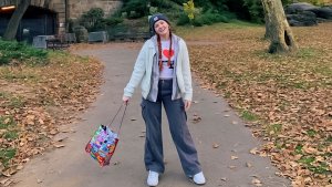


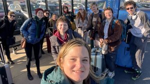


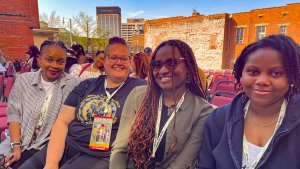


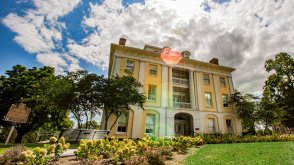
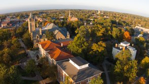
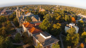
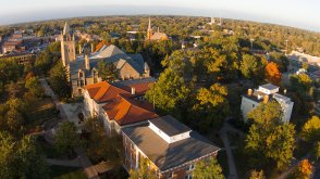
A Great Day!
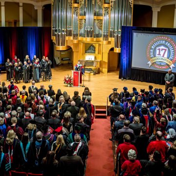
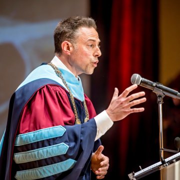
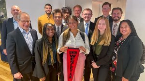
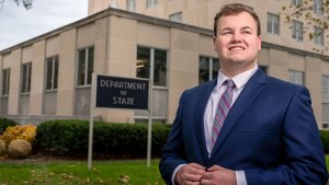
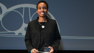
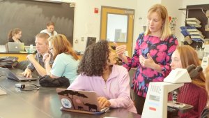
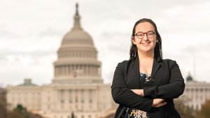
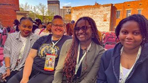
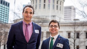
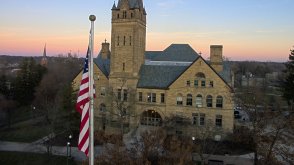
At his inauguration as OWU's 17th president, Matt vandenBerg highlighted groundbreaking initiatives, including new scholarships, partnerships, entrepreneurship competitions, and a new student hub.
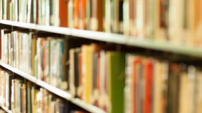
The OWU Connection
Ohio Wesleyan gives you the support, resources, and guidance to help you become who you want to be. Your OWU Connection pathway will be as unique as you are.
THINK BIG. Span academic disciplines to explore important issues and conduct mentored research with great teachers.
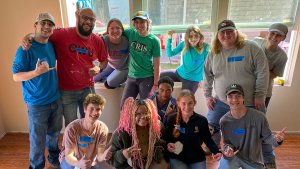
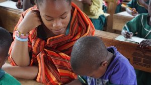
DO GOOD. Serve the community and make the world a better, more caring place.
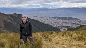
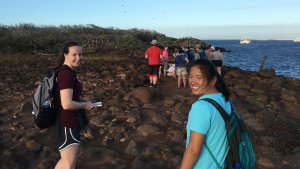
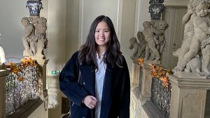
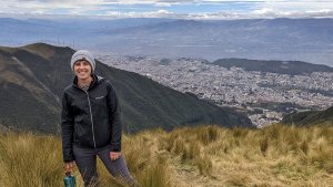
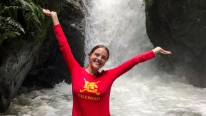
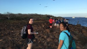
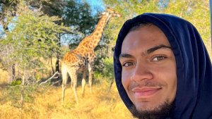
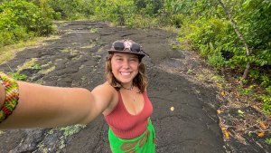
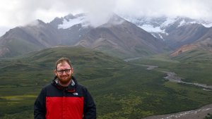
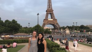
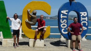
GO GLOBAL. Engage in classes and unique projects from New York to Japan, Alaska to the Galapagos Islands.
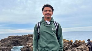
GET REAL. Connect theory to practice through internships and real-world experiences.
Learn More
OWU Connection Discovery Engine
Here's what's special about The OWU Connection: You personalize your academic program with unique Connection Experiences that combine what you care about, where you want to go, and who you want to be. Use this tool to discover some of the possibilities. Shuffle for random results or choose your pairing.
or
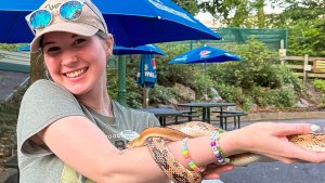
Biological Sciences
Economics & Business
Education
Physics & Astronomy
Psychology & Neuroscience
Think Big
Go Global
Get Real
Think Big
Go Global
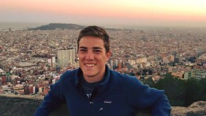
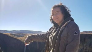
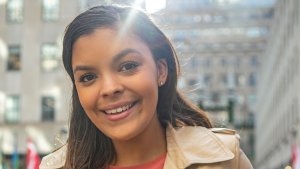
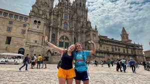
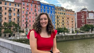
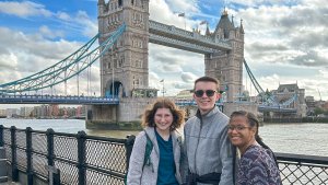
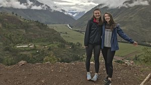
Cole Peterson ’23 traveled to the United Kingdom to conduct senior thesis research in the British Library’s India Office Archives.
History major Andrew Stock ’17 conducted original historical research to explore the career of Henry Crozier (OWU Class of 1866) as a Union artilleryman. He presented at the Student Symposium.
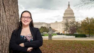
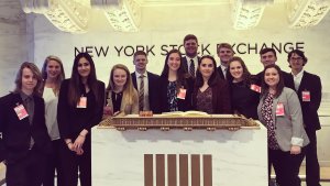
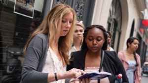
Tom Dolan '18 enjoyed a Wesleyan in Washington internship in the U.S. Senate. After he graduated, Dolan was hired as an assistant with the National Republican Senatorial Committee.
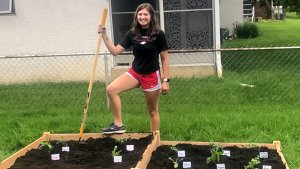
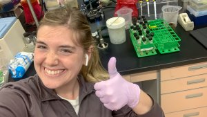
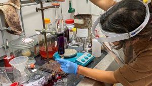
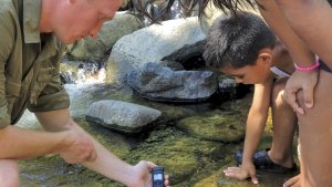
Josh Cabacungan ’24 is completing plant research in the Summer Science Research Program (SSRP).
International Business major Kellie Garvin '17 spent a semester in Barcelona, Spain, taking several fashion courses and experiencing Spanish culture.
Diego Venegas Vargas '19 spent a summer studying neutrino physics with Alfredo Galindo-Urribari of the Physics Division of Oak Ridge National Laboratory in Tennessee.
Students travel to Siena and Florence, Italy, as part of a Travel-Learning Course designed to explore how art and thought in the Renaissance intersect to form a unique ‘cultural and intellectual geography.’
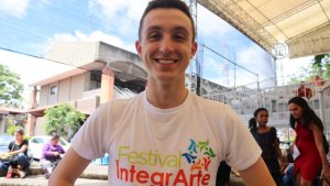
Dyna Bresson ’24 spent a semester enrolled in the New York Arts Program, during which she completed an internship with an award-winning company founded as a multi-racial ensemble dedicated to using theater as an agent for positive social change.
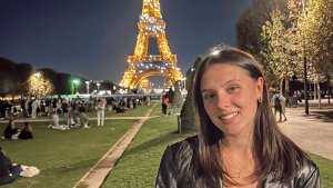
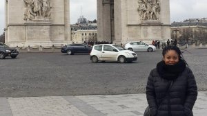
Paris Norman ’20 participated in an OWU Connection grant project to explore Pan-Africanism and its legacies in Ghana and another OWU Connection project to study French imperialism in Paris and southern France.
Raissa Kanku '19 interned with the Borgen Project, a nonprofit that aims to reduce global poverty by advocating for U.S. congressional leaders to support bills tied to foreign aid and poverty.
During her internship at the National Archives, Mackenzie Sommers '16 accessed previously classified documents for her senior project on Clara Maas, an Army nurse during the Spanish American War.
Dylan Hays '20 created a cohort of first-generation college students in the OWU Class of 2023 that met regularly to discuss the challenges and triumphs of being first-gen.
Khanh Le '16 investigated a problem in quantum physics, exploring three approximations of the energy eigenvalues of the solutions of the one-dimensional time-independent Schrodinger equation.
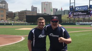
Through the Summer on the Cuyahoga program, Mic Kreutzer ’22 worked at College Now as an AmeriCorps program intern.
Quantitative Economics major Haris Ali ’22 interned with Chicago-based Upkey Corp., performing market research and providing marketing and sales support.
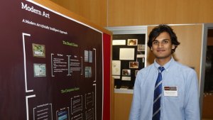
In a summer research project, Computer Science major Love Ayinde of Ilorin, Kwara, Nigeria, completed “Artificial Intelligence for Battle Line,” mentored by Prof. Sean McCulloch.
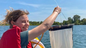
Colin Hawes ’20, Becca Porter ’20, and Katie Vonderembse ’19 worked with professor Amy Downing to study salt pollution in freshwater near campus, as part of a global research project with universities around the world.
Samuel Stull '17 interned at Nationwide Children's hospital, leading to a research project with Prof. Kira Bailey on hepatitis C and opioid abuse.
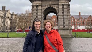
Megha Malik ’21 received a $5,000 Gilman Scholarship to travel to Dublin, Ireland, to participate in a Summer STEM Research program at University College Dublin.
Working with Prof. Scott Linder, Mathematics major Hanna Cao ’22 researched the impact of incorrectly ignoring the presence of censoring, with a goal to improve estimation methods in accounting.
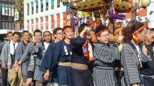
Carlie Braden ’18, Audrey Castaneda Walker ’18, Troy Jones ’17, and Kevin Rossi ’17 traveled to Japan with OWU faculty to study sustainability.
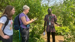
In a travel-learning course, students examined economic structures, medical systems, and West African history. During the two-week trip to Ghana, the class volunteered at a school and an orphanage.
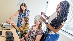
Erika Shultz '18 measured the brain waves of subjects while they completed tasks to support the theory that, depending on our goals, our brain activity switches between proactive and reactive control.
In an independent study project, Dilara Gingerich ’19 explored how the criminal justice system can use information about psychopathy to protect society from criminals with that condition.
Hien Mai '22 and Brad Orzolek '20 researched improving cognition and brain function through video games under the mentorship of Prof. Kira Bailey.
Daniel Haygood '18 worked as a summer legal intern at the County of Plumas District Attorney's Office in California.
Using a human rights framework suggested by philosopher Johnathan Wolff, Valentina Marginean ’16 found if stakeholders perceive an obligation to protect intellectual property, they risk inhibiting access to generic medicines.
Creative Writing major Kaitie Welch '20 traveled to Pordenone, Italy, to participate in an Italian-to-English translation project for a new comics museum, Palazzo Arti Fumetto Friuli.
Tiffany Moore ’20 interned at Atlantic Pictures and Breakthru Films in New York City, and she spent four weeks in New Zealand learning how to be a producer for a short film.
Business Administration major Reese Little ’22 launched the entrepreneurial venture 420Coin, a community-driven cryptocurrency created to benefit a friend paralyzed in a skiing accident.
Genetics major Cindy Hyunh ’19 and Neuroscience major Mollie Marshall ’19 received OWU Connection funding to research the effects of prenatal alcohol and methamphetamine exposure on infants in New Zealand.
In a mentored research project, Communication major My Ta ’21 explored visual meanings and variations in illustrations of 'Alice in Wonderland' from the Edwardian era.
The travel-learning Space Exploration class traveled to England and Germany, including a tour of the European Space Agency's Centre for Space Applications and Telecommunications.
S.K. Bulander ’23 created the OWU Recycling Connection, a way to help residents of the OWU and Delaware communities quickly determine what materials can be recycled locally and where.
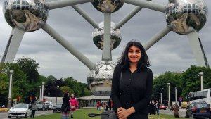
Erica Shah ’16 used an OWU Connection grant to travel to European Union headquarters in Belgium to study how the EU sustains its aims with its cultural, economic, and political diversity.
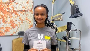
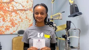
Microbiology major Gretchen Weaver ’20 worked with doctors at the Cleveland Clinic Cole Eye Institute-Louis Stokes VA Medical Center to study the effects of diabetic retinopathy.
Students visited Italy as part of a Chemistry and Art travel course, learning how chemistry plays a role in the creation, preservation, restoration, destruction, forgery detection, and analysis of art.
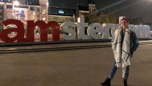
While studying abroad in Amsterdam, Alix Templeman '19 took classes on comparative sexuality studies and expanded her WGS minor into a second major.
S.K. Bulander ’23 created the OWU Recycling Connection, a way to help residents of the OWU and Delaware communities quickly determine what materials can be recycled locally and where.
Chemistry and Black World Studies major Jade Giordani '17 researched water quality and the lifestyle of the Maasai. After graduating, she became a chemistry lab technician at Battelle.
Robert Wu '20 earned a Freeman Award for Study in Asia to support a year-long experience studying at Waseda University in Tokyo and interning at a Buddhist monastery.
Communication major Sisi Fish ’24 interns at United Airlines in Chicago, Illinois.
For her Journalism studies, Gopika Nair '18 used an OWU Connection grant to explore freedom of press in Oslo, Norway. On campus, she was editor of The Transcript and interned with University Communications.
Working with Prof. Paul Dean, five sociology students surveyed about 150 OWU students to understand the factors that lead students to become involved in social activism.
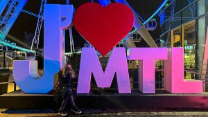
English Literature & Creative Writing major Lily Callander ’22 used an OWU Connection grant to travel to Montreal to write personal narratives that explore navigating language differences in foreign countries.
Economics and Psychology major Shaaref Shah ’17 interned with Bank of America in NYC as a Human Capital Summer Analyst. He attended grad school at the University of Pennsylvania.
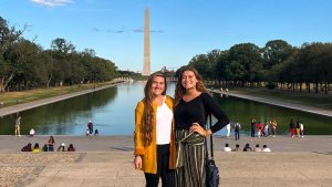
During her Wesleyan in Washington semester, Kennedy Sattler '20 interned at Airlines for America, an advocacy organization for the U.S. airline industry.
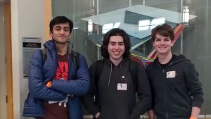
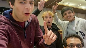
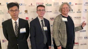
Rachel Leslie ’23 and Nick Mankowski ’25 participated in the OWU Programming Contest, an annual event for local colleges in the area.
Astrophysics and Politics & Government major Guillermo Gutierrez '18 served as an intern with the Radiant Earth Foundation and the American Physical Society.
Dyna Bresson ’24 spent a semester enrolled in the New York Arts Program, during which she completed an internship with an award-winning company founded as a multi-racial ensemble dedicated to using theater as an agent for positive social change.
Isabel Cherry ’18 worked as a youth empowerment intern at One Heart Source in Cape Town, South Africa, a nonprofit to empower young people through quality education and care.
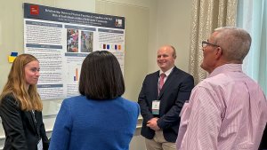
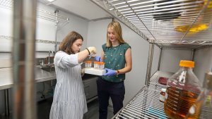
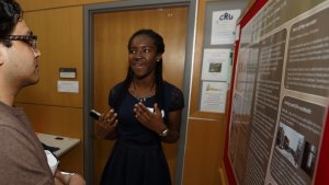
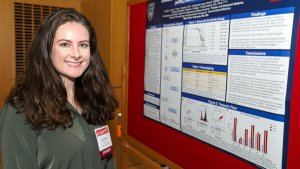
Audrey Calvin ’23, a chemistry and pre-medicine double-major, investigated proteins in a 10-week project. She presented results this fall during OWU’s annual Summer Science Research Symposium.
Cassandra Oberle ’18 conducted a research project testing leg strength and power in athletes with and without ACL reconstruction to better determine when athletes should return to training.
Diana Muzina '17 earned Departmental and University Honors for her senior research thesis exploring stereotypes and perceptions of Greek women.
Prof. Eva Paris-Huesca mentored three students who researched the Spanish Civil War and the influence of early-20th century literary and artistic movements in Federico Garcia Lorca's 1933 play "Bodas de Sangre."
In the OWU Career Connection office, job experts in education and other fields, known as career catalysts, work with students to identify opportunities and land jobs.
Under Prof. Richelle Schrock, Carson Shaw ’18 researched and presented how healthcare and culture impact cost and experience of sex reassignment surgery in Europe.
Annabel Benes ’24 is spending fall semester studying overseas in Denmark and England through the Open Campus Block Program offered by the Council on International Educational Exchange (CIEE).
Six zoology students working with Prof. Shala Hankison found that recently mated female sailfin mollies tended to produce more offspring than did females using sperm storage.
Computer Science and Data Analytics double-major David Shakarashvili ’24 interns as a data analyst with HeartWorks, Inc.
Genetics major Jack Mauter ’21 researched the effect of house dust mites on human epithelial cells at the Nationwide Children’s Hospital Department of Allergy and Immunology.
English and Communication major Dmitri Ashakih ’22 interned at the nonprofit Summer on the Cuyahoga organization as an assistant program coordinator.
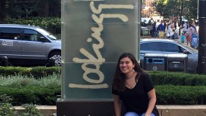
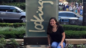
Adriana Pamela Rodriguez ’18 was a summer intern at the Lookingglass Theatre in Chicago, a nationwide leader in creating and presenting cutting-edge theatrical works.
Callia Barwick ’24 and several other members of the OWU community participated in an OWU Connection Spring Break Interfaith Service Team experience at Lakota Nation, South Dakota, for a week of “Building and Rebuilding.”
Ohio Wesleyan students visit Italy to study “From Early Museum to Contemporary Biennial” using an OWU Connection grant. They travel with mentor Erin Fletcher, director of the Ross Art Museum.
Selam Weldu ’20 received funds to attend the two-week Impact Fellowship program, which teaches college students nationwide the skills to become successful social entrepreneurs.
Adrienne Wentling ’22 used an OWU Connection grant to travel to Montreal, Québec, Canada, to conduct research on identity in French-speaking cultures.
Amanda Barry ’17 and faculty mentor Ali Skandor traveled to Tanzania for two weeks to explore memory impairment in rural Maasai communities.
Tiyinoluwa Olushola-Alao ’23 was one of several students to present original pieces at the Composition Studio Recital, performed by OWU students and faculty.
Environmental Science major Jakob Woodside '20 worked with Prof. Laurie Anderson to monitor sugar maples and their sap to determine how individual trees would change as the climate changed around them.
Communication major Sisi Fish ’24 interns at United Airlines in Chicago, Illinois.
Environmental Studies major Emily Howald ’18 helped develop OWU's Sustainability Plan, documenting sustainability initiatives across the university and serving on the Sustainability Committee.
Jackie Everetts '18 spent two weeks exploring L'Abri communities, where individuals are invited “to seek answers to honest questions about God and the significance of human life.”
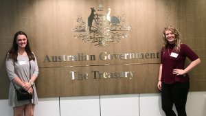
Finance Economics major Aakash Gupta ’21 interned with the Federal Deposit Insurance Corporation as part of its Financial Management Scholars Program. He worked in Risk Management Supervision.
Chemistry minor Megha Malik ’21 received a $5,000 Gilman Scholarship to travel to Dublin, Ireland, to participate in a Summer STEM Research program at University College Dublin.
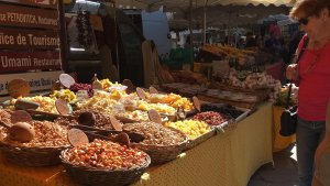
Prof. Christopher Fink traveled to Italy with two students to explore the structure, dynamics, and impact on quality of life of short food supply chains in Italy and the United States.
Blake Johnson ’25 interned with The Salvation Army and participated in an educational program in Washington, D.C., developing leadership skills, learning about hyperpartisanship, and sustaining the efforts of nonprofits.
Communication major Ash Moen ’21 traveled from Lake Erie to Hocking Hills taking photos and writing poems, which were collected in a book to encourage people to think about care about environmental issues.
Michael Barr '18 spent a year creating an honors thesis about subjectivity in James Joyce's "Ulysses" and presented at the Student Symposium.
Sophia Ahmed ’20 completed internships at Citi Bank headquarters in Texas and at technology company Thirstie in New York. Thirstie hired her after graduation.
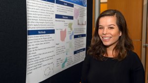
At the Student Symposium, Rebecca Penrod ’18 presented research exploring the relationship between educational outcomes and socioeconomic differences.
Dhruv Sekhawat ’26 and Inesh Tickoo ’26 participated in the 2022 HackOHI/O event, a 24-hour innovation and coding marathon held at The Ohio State University.
A group of OWU students travel to Italy to learn how chemistry is used to preserve, restore, detect forgeries, and otherwise support art as part of a Travel-Learning Course.
Ben Arnold '20 participated in a Summer Research Fellowship in the Department of Medicine at the University of Colorado Anschutz Medical Center.
Computer Science major Lan Nguyen ’18 completed two summer internships at Facebook's headquarters in Menlo Park, California. When Lan graduated, Facebook hired him as a software engineer.
Chemistry and Art major Rachel Spotts '17 used an OWU Connection grant to complete an internship at the Umlauf Sculpture Garden and Museum in Austin, Texas.
A travel-learning course studies concepts of elegance and brutality in Japanese literature before traveling to Japan.
Every year, OWU welcomes outstanding new students into the Education Fellows Program, where they enjoy scholarships and access to special career-related opportunities.
Allie Eynon '19 and Shelby Quade '18 developed materials for an online test that measures the knowledge students from 1992 to 2017 retained from an OWU Quantitative Methods course.
Dyna Bresson ’24 spent a semester enrolled in the New York Arts Program, during which she completed an internship with an award-winning company founded as a multi-racial ensemble dedicated to using theater as an agent for positive social change.
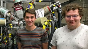
Dexter Allen ’21 researched “Intruder States and Transition Strengths in 73 As” in summer research that included work using Florida State University’s particle accelerator.
Callia Barwick ’24 and several other members of the OWU community worked with the Lakota Youth Development nonprofit as part of an OWU Connection Spring Break Interfaith Service Team to “reclaim Lakota language, culture, and spirituality.”
Diego Venegas Vargas '19 explored the mostly unknown world of nuclear physics, studying a neutron-rich isotope of gallium to better predict nuclear behavior.
As a first-year student, Alexander Sanchez '20 presented at the Student Symposium his research on Ana de Mendoza and her role as a mother in the Court of Phillip II of Spain.
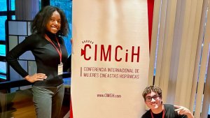
Chris MacDonald ’16 interviewed Spanish theater professionals on the role of Hispanic classical theater in Spain. He then created a documentary on the topic.
Kaytlin Ward ’21 worked with two faculty on a summer research project to find an effective route of synthesizing an organic molecule called a dipyrrin, which can be used in cancer treatment.
Students in Prof. Mary Anne Lewis Cusato’s ‘Fourteen Kilometers’ class on immigration between Africa and Europe visited a Central African refugee living in Columbus to learn his story firsthand, in French.
Brianna Robinson ’15 worked side by side with Opera Columbus, Columbus Dance Theatre, Central Ohio Symphony, and the Columbus Symphony Orchestra.
HannahJo Grimes '20 used an OWU Connection grant to compare racism in America to sectarianism in Northern Ireland.
Derek Shank '18 spent a summer at Notre Dame University working with a program that uses collected stellar data to perform a series of tests to determine a star’s properties.
Microbiology major Ashley McCracken ’19 worked with Matthew Dietz, M.D., at the West Virginia University School of Medicine to research the relationship among joint implants, infections, and treatments.
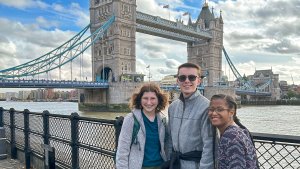
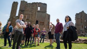
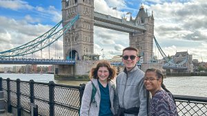
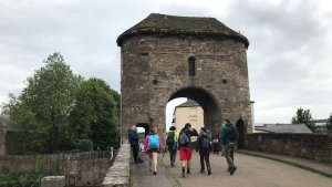
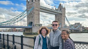
Students in the "Re-Placing Great Britain" travel course study museum narrative, architecture, theatre, poetry, and novels in the classroom and in London, Liverpool, and Manchester.
While at OWU, Rhiannon Herbert '16 held three law internships: Delaware County CASA; the Delaware, Ohio, Common Pleas Court; and a private law office.
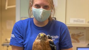
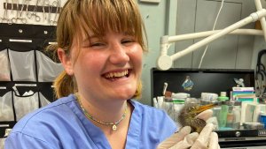
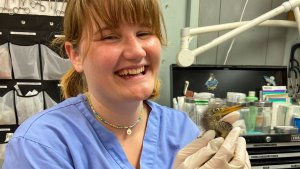
Caitlin Hyatt ’22 earned a competitive Theory-to-Practice Grant (TPG), part of the OWU Connection, to support a three-month summer internship with Pelican Harbor Seabird Station, a nonprofit wildlife rescue center located in Miami, Florida.
Psychology and French major Emma Beale '17 used an OWU Connection grant to study terrorism in France from multiple angles, including the role of propaganda and media coverage.
Sports and Exercise Management major Paddy Colligan ’24 interns as an operations assistant for the Columbus Crew.
Josh Cabacungan ’24 is completing plant research in the Summer Science Research Program (SSRP).
Psychology and Nutrition major Abby Bowman '20 used an OWU Connection grant to travel to Umbria, Italy, to explore lifestyles and diets in areas where people tend to live longer.
Accounting & Finance major Jacob Dodds ’22 had internships at Louisiana Department of Revenue, Worthington Industries, & Schneider Downs before choosing a career at Schneider Downs in Pittsburgh.
Students in Prof. Mary Anne Lewis Cusato’s "Fourteen Kilometers" class on immigration between Africa and Europe visited a Central African refugee living in Columbus to hear his story firsthand, in French.
Working with two professors, Environmental Studies major Katie Vonderembse ’19 created maps showing the distribution of Xylopia, a genus of tropical flowering plants.
Politics & Government and Astrophysics double-major Guillo Gutierrez '18 completed a senior research project on identifying influencers in social networks.
Neuroscience & Psychology major Cara Harris ’19 used an OWU Connection grant study the impact of neuroscience on every aspect of patients’ health at a public hospital in Uruguay.
Jackie Everetts '18 spent two weeks exploring L'Abri communities, where individuals are invited “to seek answers to honest questions about God and the significance of human life.”
Rachel Leslie ’23 spent a semester abroad in Quito, Ecuador, studying Latin American identity and cultural syncretism.
Cole Peterson ’23 traveled to the United Kingdom to conduct senior thesis research in the British Library’s India Office Archives.
After receiving an OWU Connection Theory-to-Practice Grant, Camy Dodd ’23 traveled to Chicago to observe professional rehearsals and make connections with theatre artists.
Kiersten Payne '17 worked with Prof. Susan Gunasti to examine the language contemporaries used to discuss the Donatists, beginning in the 4th century, and Anabaptists, beginning in the 16th century.
During a summer internship at the Mayo Clinic, Eric Baughman '17 participated in a research laboratory examining congenital heart diseases using stem cells.
For two summers, Jack Mauter ’21 conducted research on respiratory conditions with a doctor in the Nationwide Children’s Hospital Department of Allergy and Immunology.
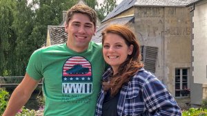
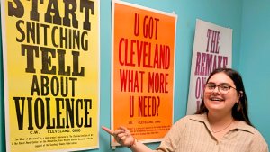
Sisi Fish ’24 is spending the summer living and working in the Cleveland, Ohio, area, where she is serving as the marketing and special projects intern for the Summer on the Cuyahoga (SOTC) organization.
At the OWU Student Symposium, three students presented their research on the representation of women in francophone films, portrayals that challenged or confirmed stereotypes.
At the Student Symposium, Cierra Cresanto ’17 presented her research into why some regimes were overthrown in the Arab Spring and others were not.
Exercise Science major Emily Sheridan ’21 worked with two faculty members on a research project to examine the effectiveness of yoga in reducing anxiety in children and adolescents.
Working with Prof. Robert Harmon, Alec Martin '19 observed the starspots of a young star in order to better understand how a star's magnetic fields are created.
In the travel course “Mexican Migration Experience,” students explored the pyramids of Teotihuacan and stayed with families to become more familiar with Mexican culture.
Philosophy & Psychology major Max Johnson ’21 used an OWU Connection grant to complete a 200-hour training course to earn yoga teacher certification through the Yoga Alliance.
Pre-Medicine major Alex Goldenberg ’25 founds nonprofit to provide clothing and shoes to Delaware County homeless community.
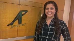
Film Studies major Leah Crawford ’19 spent a summer in Culver City and Beverly Hills, California, completing internships with Grey Matter Productions and Artists First.
Through the Summer on the Cuyahoga program, Women's & Gender Studies major Mic Kreutzer ’22 worked at College Now as an AmeriCorps program intern.
Computer Science major Eugene Kramskoi '19 worked with Prof. Sean McCulloch and a student from Syracuse University in a summer project developing a computer program to play the board game Pandemic.
Sports & Exercise Management major Zack Katona '17 used skills from his Great Lakes Summer Internship to enhance his entrepreneurial efforts to launch a dietary supplement business called Rise.
International Studies major Jessica Sanford '17 interned as a political coordinator at the National Republican Senatorial Committee in Washington, D.C.
Rachel Tallmadge ’14 conducted research on human trafficking. Later, as a law student at Ohio State, she received a fellowship to work for a year with juvenile victims of human trafficking.
In a senior research project, Middle Childhood Education major Natalie French ’17 explored LGBT representation in young adult literature.
Nick Mankowski ’25 has been participating in an internship with 3E since mid-July under the supervision of two of the company’s senior software engineers.
Brianna DeMuth '23 worked with Prof. James Franklin on research into protest waves in various countries that have led to some kind of accountability or regime change.
Mackenzie Sommers ’16 helped digitize a collection of Confederate records as a part of her internship duties at the National Archives via the Wesleyan in Washington program.
Samantha Merino ’21 spent a semester working remotely as the Spanish interpretation and translation intern for the Canton, Ohio-based Immigrant Worker Project.
Economics major Max Beard ’22 interned at the Stanford Graduate School of Business, finding, summarizing, and analyzing research for a literature review of e-payment technologies in developing countries.
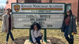
Emma Tarawally '20, Raissa Kanku '19, and Eva Churu '19 traveled to Chicago to examine the global Afrocentric education model of Kwame Nkrumah Academy, a charter school in Chicago.
In a senior research project, Harrison Nickels ’19 explored the influence of Taoist philosophy on Chinese art that reflected a passive approach to the progression of mundane events.
Prof. Christopher Fink has traveled to Italy with more than 50 students to explore food studies.
Colin Hawes ’20, Becca Porter ’20, and Katie Vonderembse ’19 worked with professor Amy Downing to study salt pollution in freshwater near campus, as part of a global research project with universities around the world.
Dyna Bresson ’24 spent a semester enrolled in the New York Arts Program, during which she completed an internship with an award-winning company founded as a multi-racial ensemble dedicated to using theater as an agent for positive social change.
In a summer internship, Isa Johnson ’22 worked in plant genetics as part of the cutting-edge Plant Genome Research Program at Cornell University.
Michael Heeschen '20 participated in the Summer Science Research Program, where he studied fundamental properties of heavier nuclei, such as gallium.
Peyton Hardesty ’20 worked as an apprentice with the head apiarist at Stratford Ecological Center researching healthy hive management and observing honey bees’ natural processes.
S.K. Bulander ’23 created the OWU Recycling Connection, a way to help residents of the OWU and Delaware communities quickly determine what materials can be recycled locally and where.
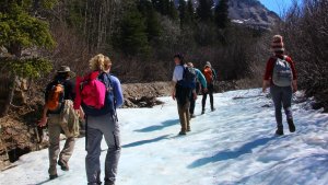
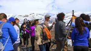
Two travel courses, “Mathematical Models of Climate” and “Plant Responses to Global Change,” have completed their semester with research and study in Alaska.
Erin Cannon '18 and Caitlin Maggio '17 used an OWU Connection grant to study the impact of international accounting standards in Australia.
Rachel Leslie ’23 spent a semester abroad in Quito, Ecuador, studying Latin American identity and cultural syncretism.
Biochemistry major Madeleine Sorrick '19 spent a summer working with two professors at The Ohio State University exploring "Yeast Display of Cysteine Free 3E8 Antibodies."
Accounting and International Studies major Ahmed Hamed ’20 earned a Gilman International Scholarship from the U.S. Department of State to study Arabic in Morocco.
Business Administration major Avianna Carmoega ’20 interned with the nonprofit Human Connections in Bucerías, Mexico, working on a variety of marketing projects.
For her senior thesis, Amanda Hays ’20 completed a two-semester honors project in which she took an in-depth look at the relationship between Leo Tolstoy and Mahatma Gandhi.
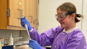
Abby Biddle ’23 is participating in Ohio Wesleyan’s annual Summer Science Research Program (SSRP), researching anxiety and mood disorders and the impact of stress during adolescence.
In summer research with Prof. Dustin Reichard, Joe Brush '20 studied aggression in two male songbird species.
Emily Feldmesser ’16 worked as an intern at The Brookings Institution, one of the world’s foremost think tanks, as part of a Wesleyan in Washington experience.
Creative Writing major Sarah Jonassen ’22 completed a summer internship with Franklin County’s Mifflin Township, expanding her writing portfolio.
Karli Walsh ’23 traveled to London with her professor over October’s mid-semester break to research and compare food allergen regulations in the United Kingdom and the United States.
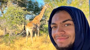
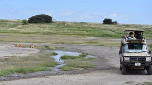
Amanda Marshall ’16 used an OWU grant to help a research team at the Karongwe Private Game Reserve in South Africa collect data on cheetahs, lions, elephants, leopards, and hyenas.
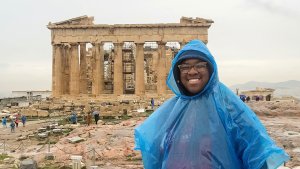
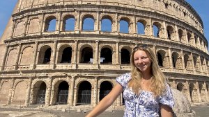
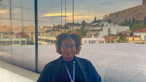
During a semester or summer, students participate in programs such as the American School of Classical Studies at Athens, the College Year in Athens, or the Institute for Roman Culture in Rome.
Catie Kocian '18 and James Ormerod '17 traveled to Japan & Taiwan to study waste management. Their trip was funded by a Luce Grant.
Early Childhood Education majors Ali Phillips ’16 and Whitney Weadock ’16 received an OWU grant to attend a global childhood education summit in San Jose, Costa Rica.
Business major Robert Wu ’20 earned a $7,000 Freeman Award for Study in Asia to support studying abroad for a year at Waseda University in Tokyo, Japan.
Philosophy minor Samantha Merino ’21 spent a semester working remotely as the Spanish interpretation and translation intern for the Canton, Ohio-based Immigrant Worker Project.
After receiving an OWU Connection Theory-to-Practice Grant, Camy Dodd ’23 traveled to Chicago to observe professional rehearsals and make connections with theatre artists.
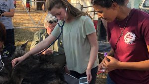
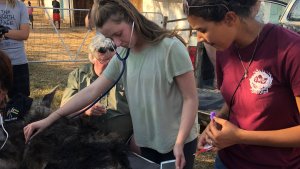
Lauren Kiebler ’16 completed several internships and gained valuable veterinary experience at Severna Park Veterinary Hospital, Mobile Pet Vet, and the Humane Society of Delaware County.
S.K. Bulander ’23 created the OWU Recycling Connection, a way to help residents of the OWU and Delaware communities quickly determine what materials can be recycled locally and where.
Taimur Iftikhar '20 and Avianna Carmoega ’20 interned with Human Connections, a nonprofit social enterprise based in Bucerías, Mexico.
Bella Campos Hintzman ’22 used OWU Connection grant funding to conduct research in California in support of her senior capstone project in regional theater career preparation.
Inesh Tickoo ’26 participated in the 2022 HackOHI/O event, a 24-hour innovation and coding marathon held at The Ohio State University.
Kristen Krak ’15 used an OWU Connection grant to attend the National Theatre Institute summer intensive in Waterford, Connecticut, for classes, collaborative creation, and workshops.
Zoology major Becca Strider ’20 received a full-tuition program scholarship from the Council on International Educational Exchange to study in Central America for 18 weeks.
Erin Fannin ’18 and Karis Lowrie ’18 received OWU’s Herbst Prize to fund their study in Rome, Florence, and Pompeii.
Eli Reed '19 used an OWU Connection grant to compare sectarianism in Northern Ireland to American race relations.
Aadarsha Gopala Reddy ’23 participated in OWU’s annual Summer Science Research Program (SSRP), researching the use of artificial intelligence as it applies to modern board games.
Communication majors Erin Ross ’21 and Jacey Scheffel ’21 filmed and produced a documentary on Delaware County’s annual Olentangy River Cleanup.
In the travel course "The Literary Politics of Ireland," students attended Listowel Writer's Week, a weeklong literary festival celebrating Irish writing, theatre, and film.
Bulbul Bhati ’23 completed a data analytics internship for RxNXT, an Ohio-based company that provides software to enable employees to search for the best available price on medications.
Through the Summer on the Cuyahoga program, Samavi Sultana ’22, from Baltimore, Maryland, worked as an intern at Vecmar, a computer solutions firm.
Botany and Biochemistry double-major Carly Sanders ’24 participated in OWU’s annual Summer Science Research Program (SSRP), studying under the mentorship of OWU professor Chris Wolverton.
Michael Heeschen ’20 presented his research, “Level Scheme and Spin Assignments in 70Ga,” at the Student Symposium.
Students of this travel-learning course traveled to New Orleans for spring break to learn about traditional music of the region and the roots of modern styles like rock and jazz.
Rachel Leslie ’23 participated in the OWU Programming Contest, an annual event for local colleges in the area.
After receiving an OWU Connection Theory-to-Practice Grant, Camy Dodd ’23 traveled to Chicago to observe professional rehearsals and make connections with theatre artists.
Theatre major Jasmine Lew ’22 gained student-teaching experience at the nonprofit Chicago Center for Urban Life and Culture.
At Washington University School of Medicine in St. Louis, Biochemistry & Neuroscience major Hien Ngoc Mai ’22 worked on research to develop biomarkers with radioactive chains to diagnose neurodegenerative diseases.
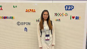
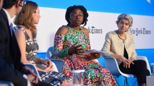
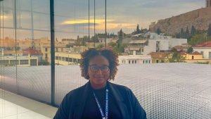
Ellie Bearss ’22 was one of 24 students nationwide to participate in the NY Times Athens Democracy Forum. Her video was played at a panel discussion on Art as Activism.
Business Administration major Aidan Stout '19 received a grant to support his summer in Boulder, Colorado, interning with Bob Gordon '88 of Berkshire Hathaway HomeServices.
Daniel Eilert ’23 conducts neuroimaging research at Nationwide Children’s Hospital.
Prof. Jason Hiester and seven students received an OWU Connection grant to travel to Austria to participate in workshops and masterclasses and attend the Salzburg Music Festival.
In a Summer Science Research Project, Aidan Shumaker ’20 worked with Prof. Laurie Anderson to research greenhouse gas emissions underneath invasive honeysuckle and native spicebush shrub species.
In a directed reading project on women and power in early modern Spain, Maria Alonso ’20 worked with Prof. Glenda Nieto Cubas to explore the enigma behind Joanna of Castile’s mental state.
In a project with Prof. Eva Paris-Huesca, Landry Cowles ’20 and Gretchen Weaver ’20 explored how health literacy competency is dependent upon language competency and cultural impact.
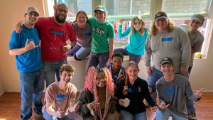
In a spring break service trip, a team of OWU students volunteered to help rebuild a family's Houston-area home following Hurricane Harvey.
Bavneet Singh ’25 recently completed a 13-week remote internship with Movement Genius, a digital wellness platform that provides live and on-demand classes to help people improve their mental, emotional, and physical well-being.
Myles Steed ’23 conducted summer research exploring faster, more effective ways to create a chemical called a dipyrrin ligand, which improves the use of hydrogen fuel cells.
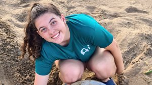
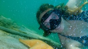
Hannah Cox ’24 participated in an OWU Connection grant-supported internship in Greece aiding loggerhead sea turtles.
For her senior capstone project, Theatre and English major Hannah Simpson ’16 explored how we “play” our gender and how the role of Hamlet changes when performed by a female.
Amanda Hays '20 used an OWU Connection grant to complete an internship at the Hermitage State Museum in St. Petersburg, the second-largest art museum in the world.
Kolby Brock ’23 and Karli Walsh ’23 traveled to London with their professor over October’s mid-semester break to research and compare food allergen regulations in the United Kingdom and the United States.
Film Studies and Spanish major Tess Meddings '22 combined two of her academic passions by serving as the intern for Ohio Wesleyan’s Hispanic Film Festival.
After receiving an OWU Connection Theory-to-Practice Grant, Camy Dodd ’23 traveled to Chicago to observe professional rehearsals and make connections with theatre artists.
Carly LoVullo ’16 received a $24,000 fellowship to support a yearlong study of best practices for community-based disease surveillance of dengue fever in three Tanzanian coastal regions.
Sarah Bergman '18, James Zoller '18, and Lauren Boedicker '18 worked with Prof. Allen Pistner to find a more affordable and effective chemical catalyst for biodegradable plastic.
Communications major and Fine Arts minor Sisi Fish ’24 spent a summer working for Cleveland’s Summer on the Cuyahoga program as a marketing and special projects intern.
Accounting major J.T. Knoble ’17 and two other students received an OWU Connection grant to study generally accepted accounting principles (GAAP) in Ireland.
Two travel courses, “Plant Responses to Global Change” and “Mathematical Models of Climate,” have completed their classes with research trips to Alaska.
As part of the Wesleyan in Washington program, Emily Feldmesser ’16 interned at the Brookings Institution, one of the world’s foremost think tanks, in Washington, D.C.
Emma Hall ’21 Hall spent a summer researching “Stress, Locomotion, and Zebrafish Mutant Analysis” under the mentorship of Karl Clark, Ph.D., at Mayo Clinic in Rochester, Minnesota.
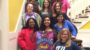
Many students intern at OWU’s Spectrum Resource Center or Women’s Resource Center, where you might organize campus events including the annual Take Back the Night march.
Cierra Cresanto '17 worked with Prof. James Franklin on research into why some regimes were overthrown in the Arab Spring and others were not. She presented at a professional conference.
Communication and English major Dmitri Ashakih ’22 interned at the nonprofit Summer on the Cuyahoga organization as an assistant program coordinator.
In a senior research project, Katie Kuckelheim ’19 explored why women in student leadership may change their communication style based on the gender makeup of a group.
Chloe Sullivan ’24 and several fellow OWU students spent the summer working to complete NASA-funded research in the laboratory of Chris Wolverton, Ph.D.
Katherine Berger '16 completed an internship in the Ohio Attorney General's office. After graduating, she moved on to The Ohio State University Moritz College of Law.
Health Promotion major Lauren Mussenden ’22 and Nutrition major Leigh Stavar ’22 completed a summer internship working with the Cooking Matters community education program.
Trey Olsen ’18: "It was extremely rewarding to apply my knowledge from Ohio Wesleyan and represent OWU among an internship class from Ivy League and other prestigious universities."
Kelly Maier '14 and Emma Goetz '15 visited Sippo and Helsinki, Finland, to study the nation’s renowned education system.
Exercise Science major Jacqueline Feliciano ’18 studied muscle imbalances and performance changes during a field hockey season. She moved on to the Doctor of Physical Therapy Program at Ohio University.
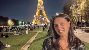
The travel-learning course “Introduction to French Literature” visited France for a week, with stops in Paris, Versailles, and Provins.
Emma Blackburn ’22 worked on summer research with Prof. Bethany Rudd to improve global climate models to better account for the impact of lake and sea spray aerosols.
For her senior thesis, Amanda Hays ’20 completed an honors project in which she explored how Leo Tolstoy’s philosophy influenced the movement for Indian independence.
Economics major Erica Shah ’16 used an OWU Connection grant to travel to European Union headquarters in Belgium to study how the EU sustains its aims with its cultural, economic, and political diversity.
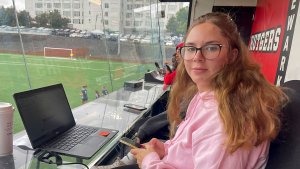
Sports & Exercise Management major Kristie Prendergast ’15 worked in sponsorship and marketing for Major League Lacrosse’s Ohio Machine, where she worked with the announcer during the Machine’s summer season.
Diego Venegas Vargas '19 spent a summer studying neutrino physics with Alfredo Galindo-Urribari of the Physics Division of Oak Ridge National Laboratory in Tennessee.
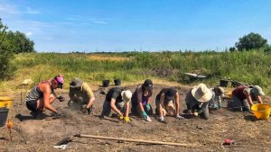
Psychology and Sociology/Anthropology major Bridget Roddy ’20 used an OWU Connection grant to serve on the volunteer excavation team at the Roman fort of Halmyris in Romania.
International Business major Emily Julius ’16 explored obstacles that Japanese women entrepreneurs face, researching successful Japanese women and conducting interviews with Japanese businesswomen.
Amanda Jewell ’19, Kaito Iwasaki ’23, and Sakshi Gupta ’22 completed 10-week summer projects studying starspots on LO Pegasi under the mentorship of Prof. Robert Harmon.
Serena George ’19 researched paternity and mating behaviors in the Sailfin Molly under Prof. Shala Hankison. Serena was admitted to the DVM program at University of Wisconsin.
Sarah Jonassen ’22 completed a summer internship with Franklin County’s Mifflin Township, expanding her writing portfolio.
Nam Tran Hoang ’16 constructed and characterized a mathematical model to assess abnormal numbers of repeating segments of DNA. He worked with Prof. Craig Jackson.
Working with two Botany professors, Katie Vonderembse ’19 created maps showing the distribution of Xylopia, a genus of tropical flowering plants.
After interning with an improv company in New York, Hannah Wargo '20 created her own sketch show and performed it for the OWU community for her senior project.
Leah Crawford ’19 spent her summer in Culver City and Beverly Hills, CA, completing internships with Grey Matter Productions and Artists First.
Julia Stone ’16 lived with a host family while she studied abroad for a semester in Aix-en-Provence.
Bella Campos Hintzman ’22 used OWU Connection grant funding to conduct research in California in support of her senior capstone project in regional theater career preparation.
Special Education major Alanna Spalsbury ’16 expanded personal experience as a one-on-one aide for a kindergartener with hyperlexia into a research project exploring the advanced reading ability.
The East Asian Politics travel-learning class traveled to Korea.
At the OWU Student Symposium, Chloe Dyer ’18 presented her research on perceptions of Irish nationalism, identity, and the role of women in the poetry of “The Lost Land” by Eavan Boland.
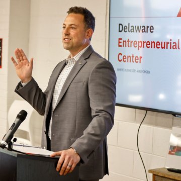
Pre-Medicine major Alex Goldenberg ’25 founds nonprofit with assistance from the Delaware Entrepreneurial Center at OWU.
Environmental Science & Zoology major Serena George ’19 conducted summer research exploring the presence of ticks on roadkill vertebrates in northern Tanzania.
Students in the Accounting Fellows program participate in at least one internship and make connections with OWU alumni working in New York and Washington, D.C.
Taimur Iftikhar '20 and Avianna Carmoega ’20 interned with Human Connections, a nonprofit social enterprise based in Bucerías, Mexico.
Film Studies and Spanish major Tess Meddings '22 combined two of her academic passions by serving as the intern for Ohio Wesleyan’s Hispanic Film Festival.
Slater Sabo '19 presented an original scholarly paper at the National Phi Alpha Theta Conference in New Orleans.
Finance Economics major Caroline Kermode '20 spent a summer interning with Charles Schwab's Richfield, Ohio, office, working in the Retirement Investment and Document Services Department.
Business Administration major Kera Bussey-Sims ’18 traveled to Seattle to interview leaders at Nordstrom and Amazon on how technology is being integrated into the fashion business.
Environmental Studies major Peyton Hardesty ’20 worked as an apprentice with the head apiarist at Stratford Ecological Center researching healthy hive management and observing honey bees’ natural processes.
Genetics major Cindy Hyunh ’19 and Neuroscience major Mollie Marshall ’19 researched the effects of prenatal alcohol and methamphetamine exposure on infants in New Zealand.
Kristen Nooney ’18, Jessica Sanford ’17, and Meg Teitelman ’18 combined creative and analytical work, reimagining a contemporary film adaptation of Flaubert's "Madame Bovary."
Early Childhood Education major Gabi Coty ’17 explored methods used around the world for teaching mathematics in her senior research project.
Pre-Medicine, Biology, and Environmental Science major Madison Cartnal ’24 was one of 12 students working on summer NASA-funded research on how plants respond to gravity.
Elizabeth Sumoza ’25 traveled to New York City and Denver to complete a research project titled “Exploring Latinx Productions of Early Modern Hispanic Texts.”
Astrophysics and Politics & Government double-major Guillo Gutierrez '18 completed a senior research project on identifying influencers in social networks.
Neuroscience and Pre-Med major Ben Arnold '20 participated in a Summer Research Fellowship in the Department of Medicine at the University of Colorado Anschutz Medical Center.
Zoology major Reagan Jennings ’23 used an OWU Connection grant to do comparative research observing wild and captive harbor seal social behaviors.
Exercise Science major Karson Stevenson ’16 traveled to Europe with an OWU Connection grant to study alcohol-induced aggression among soccer fans.
Alyssa Richter ’17 and Lauren Kibler ’16 worked with Prof. Danielle Hamill, researching worms with a mutant gene to better understand cell division.
Max Johnson ’21 used an OWU Connection grant to complete a 200-hour training course to earn yoga teacher certification through the Yoga Alliance.
Curry Carr ’21 worked with Prof. Shari Stone-Mediatore to examine arguments that facilitated the enactment of harsh U.S. sentencing laws, focusing on truth in sentencing laws in Illinois.
Environmental Science major Eva Blockstein '19 used an OWU Connection grant to volunteer at the Panama Amphibian Rescue and Conservation Center, studying spindly leg syndrome in endangered frog species.
The “Cervantes and the Quixote” travel-learning course visited Spain to make connections between literature read in class and the culture, society, history, and arts of Spain.
Selam Weldu ’20 received funds to attend the two-week Impact Fellowship program, which teaches college students nationwide the skills to become successful social entrepreneurs.
At OWU’s Student Symposium, Alyssa DiPadova '19 presented her research project, “Charisma’s Triumph over Organization: Peronism Throughout the Decades.”
Funded by an OWU grant, Carly Zelenski ’15 interned with the East Meets West Foundation in Vietnam, traveling to various provinces to conduct onsite evaluations for clean-water projects.
Zoology major Kara Smith ’20 conducted a behavioral observation study focused on the Matschie’s tree kangaroo, exploring how crouching behavior may be an intention movement for jumping.
Astrophysics and Politics & Government major Guillermo Gutierrez '18 served as an intern with the Radiant Earth Foundation and the American Physical Society.
Caitlin Hyatt ’22 earned a competitive Theory-to-Practice Grant (TPG), part of the OWU Connection, to support a three-month summer internship with Pelican Harbor Seabird Station, a nonprofit wildlife rescue center located in Miami, Florida.
Maeve Nash ’16 completed student teaching in Columbus with drama educator & OWU alum Emily Foster Whittaker. At OWU, Maeve performed, stage managed, and studied in Ireland.
The Modernity & Colonialism class traveled to Chiapas, Mexico, lived in a Zapatista community, learned about indigenous philosophies, and participated in a mural-painting project.
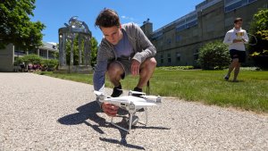
Integrated Science for Teachers major Austin Riegel ’21 used drone technology to help the community with environmental concerns and to promote citizen science in Costa Rica.
Exercise Science major Megan Sievers ’20 conducted research to help clarify the role of multiple variables in the development of shin splints in college runners.
Belle Norman ’22 participated in a Bishop Externship at Nationwide Children’s Hospital shadowing Kim Leary ’09 to learn more about Autism Spectrum Disorder.
In a Cincinnati Children’s research fellowship, Madeline Bonfield ’18 examined the correlation between the presence of a language gap and lower social function and negative behaviors in children who are deaf or hard of hearing.
Students in the travel course “Modernity & Colonialism,” spent a week with a Zapatista community in Chiapas, Mexico, reconsidering concepts such as progress, justice, and truth.
Josh Pletcher '21 presented an iCubed lecture about his time spent cataloging artifacts in OWU's Brandt Museum of Zoology.
Madeleine Rea ’20 worked with Prof. Nathan Rowley on research using satellite imaging data to explore the effects of red algae on glacier melt rates of the Harding Icefield in Alaska.
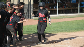
April 28
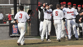
Ohio Wesleyan University Softball vs Wooster - Strike Out Cancer
1 p.m. – 3 p.m.
More Information


May 2
MH Workshop: Building Resilience During Transitions/Final Exam Stress Break
12 p.m. – 1 p.m.
More Information
Headlines
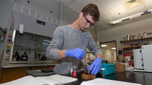
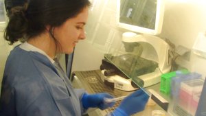
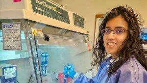
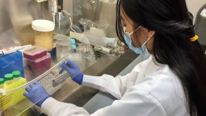
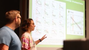
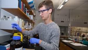
Elemental Research
Make The Connection – April 24, 2024
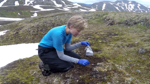
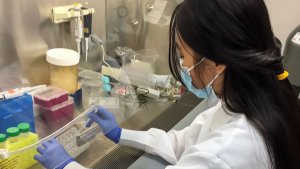
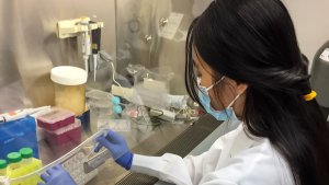
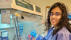
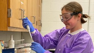
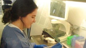
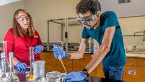
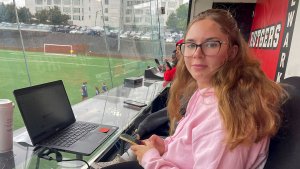
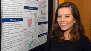
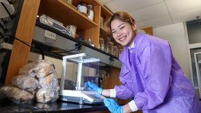
Ohio Wesleyan Student Studies the Effects of Selenium on Mushroom Growth, Development
More information
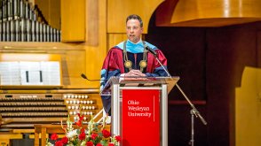
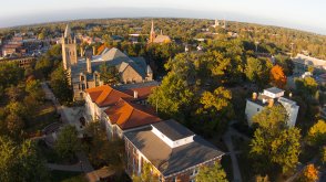
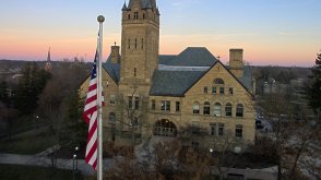
17th President
Feature Story – April 23, 2024
Ohio Wesleyan's Matt vandenBerg Announces Groundbreaking Initiatives at April 19 Inauguration
More information
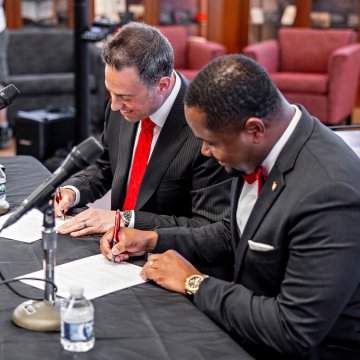
Groundbreaking Agreement
Press Release – April 18, 2024
'Landmark Partnership'
Press Release – April 18, 2024
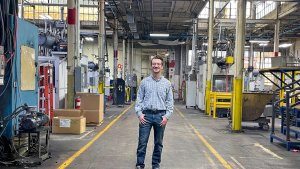
Big Business
Press Release – April 16, 2024
More News Headlines